Muscles In The Body Diagram - Solved Draw The Free Body Diagram Showing The Forces That Chegg Com
In this image, you will find frontalis, orbicularis oculi, zygomaticus, masseter, orbicularis oris, sternocleidomasteoid. We hope this post inspired you and help you what you are looking for. Below are two human body muscle diagrams, showing the front and back of the body. Anterior muscles in the body. It permits movement of the body, maintains posture and circulates blood throughout the body. There are around 650 skeletal muscles within the typical human body. Femur, capsule of knee, head of fibula. There are approximately 640 skeletal muscles within the typical human, and almost every muscle constitutes one part of a pair of identical bilateral muscles, found on both sides, resulting in approximately 320 pairs of muscles, as presented in this article. Click on the name of a muscle for a page about that muscle (works for most labels). Muscle diagram, most important muscles of an athletic black man, anterior and posterior view, male body. Heaviest muscle in body, extends/straightens leg at hip during walking.
See more ideas about body diagram, muscle anatomy, muscles in your body. First the head, then the neck, the shoulders and arms, and only then the lower parts of the body. The muscles of the spine anatomy chart shows every one of the many layers of muscle in the spine and back, using beautifully illustrated and detailed representations of the human anatomical structure. The muscular system is made up of specialized cells called muscle fibers. Feel free to browse at our anatomy categories and we hope you can find your inspiration here. Diagrams of the muscles and guide to how they work. Their main function is contractibility. There are around 650 skeletal muscles within the typical human body. This image is titled muscles of the body diagram picture and is attached to our article about 3 main muscle types in the human body.
The interactive muscle anatomy diagram shown below outlines the major superficial (i.e.
Thank you for visiting major muscles of the body diagram pictures. It permits movement of the body, maintains posture and circulates blood throughout the body. The muscles of the human body are responsible for movement; We hope this post inspired you and help you what you are looking for. Muscle diagrams are a great way to get an overview of all of the muscles within a body region. Within this group of back muscles you will find the latissimus dorsi, the trapezius, levator scapulae and the rhomboids. Each type of muscle tissue in the human smooth muscle is found in the walls of hollow organs throughout the body. See more ideas about muscle diagram, human anatomy and physiology, medical anatomy. In the diagrams below, i'll be showing muscle groups in color, with a black line to show the forms that would show through the skin (i also show protruding bones that would do the then cover it instead with a thick bathing towel. Almost every movement in the body is the outcome of muscle contraction. This image is titled muscles of the body diagram picture and is attached to our article about 3 main muscle types in the human body. Diagrams of the muscles and guide to how they work. Flexes leg at knee joint and extend thigh at hip joint tibialis anterior tibia first cuneiform and first metatarsal. Part of quadriceps group, extends leg at knee.
The muscles of the spine anatomy chart shows every one of the many layers of muscle in the spine and back, using beautifully illustrated and detailed representations of the human anatomical structure. It also helps raise the body from a supine. Flexes leg at knee joint and extend thigh at hip joint tibialis anterior tibia first cuneiform and first metatarsal. The next life study seated female figure, shows the upper part of the pectoralis major positioned flat against the rib cage, with very the muscle helps bend the torso forward in the movement known as the flexion of the vertebral column. The interactive muscle anatomy diagram shown below outlines the major superficial (i.e. There are approximately 640 skeletal muscles within the typical human, and almost every muscle constitutes one part of a pair of identical bilateral muscles, found on both sides, resulting in approximately 320 pairs of muscles, as presented in this article. Heaviest muscle in body, extends/straightens leg at hip during walking. Almost every movement in the body is the outcome of muscle contraction. The human muscular system is complex and has many functions in the body.

Almost every movement in the body is the outcome of muscle contraction.
Click on the name of a muscle for a page about that muscle (works for most labels). The muscles of the human body are responsible for movement; Diagrams of the muscles and guide to how they work. The interactive muscle anatomy diagram shown below outlines the major superficial (i.e. Muscle diagram, most important muscles of an athletic black man, anterior and posterior view, male body. Calcaneus tibial nerve plantar flexes foot. Muscles, connected to bones or internal organs and blood vessels, are in charge for movement. Muscle diagrams are a great way to get an overview of all of the muscles within a body region. It permits movement of the body, maintains posture and circulates blood throughout the body. Learn about them and what the skeletal muscles are the bulk of muscles in the body. Freetrainers.com has a vast selection of exercises which are used throughout our workout plans. If you found any images copyrighted to yours, please contact us and we.
Feel free to browse at our anatomy categories and we hope you can find your inspiration here. There are approximately 640 skeletal muscles within the typical human, and almost every muscle constitutes one part of a pair of identical bilateral muscles, found on both sides, resulting in approximately 320 pairs of muscles, as presented in this article. These include mobility, stability, posture, circulation, digestion, and more.
Human muscle system, the muscles of the human body that work the skeletal system, that are under voluntary control, and that are concerned with the following sections provide a basic framework for the understanding of gross human muscular anatomy, with descriptions of the large muscle groups.
Feel free to browse at our anatomy categories and we hope you can find your inspiration here. There are approximately 640 skeletal muscles within the typical human, and almost every muscle constitutes one part of a pair of identical bilateral muscles, found on both sides, resulting in approximately 320 pairs of muscles, as presented in this article. But, your soleus muscle in your lower leg and muscles in your back involved in maintaining posture contain mainly slow twitch muscle fibres. There are around 650 skeletal muscles within the typical human body. This is a table of skeletal muscles of the human anatomy. It should be noted that there are many more muscles in the body that are not addressed by this muscle anatomy diagram. The muscles of the spine anatomy chart shows every one of the many layers of muscle in the spine and back, using beautifully illustrated and detailed representations of the human anatomical structure. Calcaneus tibial nerve plantar flexes foot. If you found any images copyrighted to yours, please contact us and we. Use the location, shape and surrounding structures to help you.

See more ideas about body diagram, muscle anatomy, muscles in your body.

The muscles of the human body are responsible for movement;

It should be noted that there are many more muscles in the body that are not addressed by this muscle anatomy diagram.

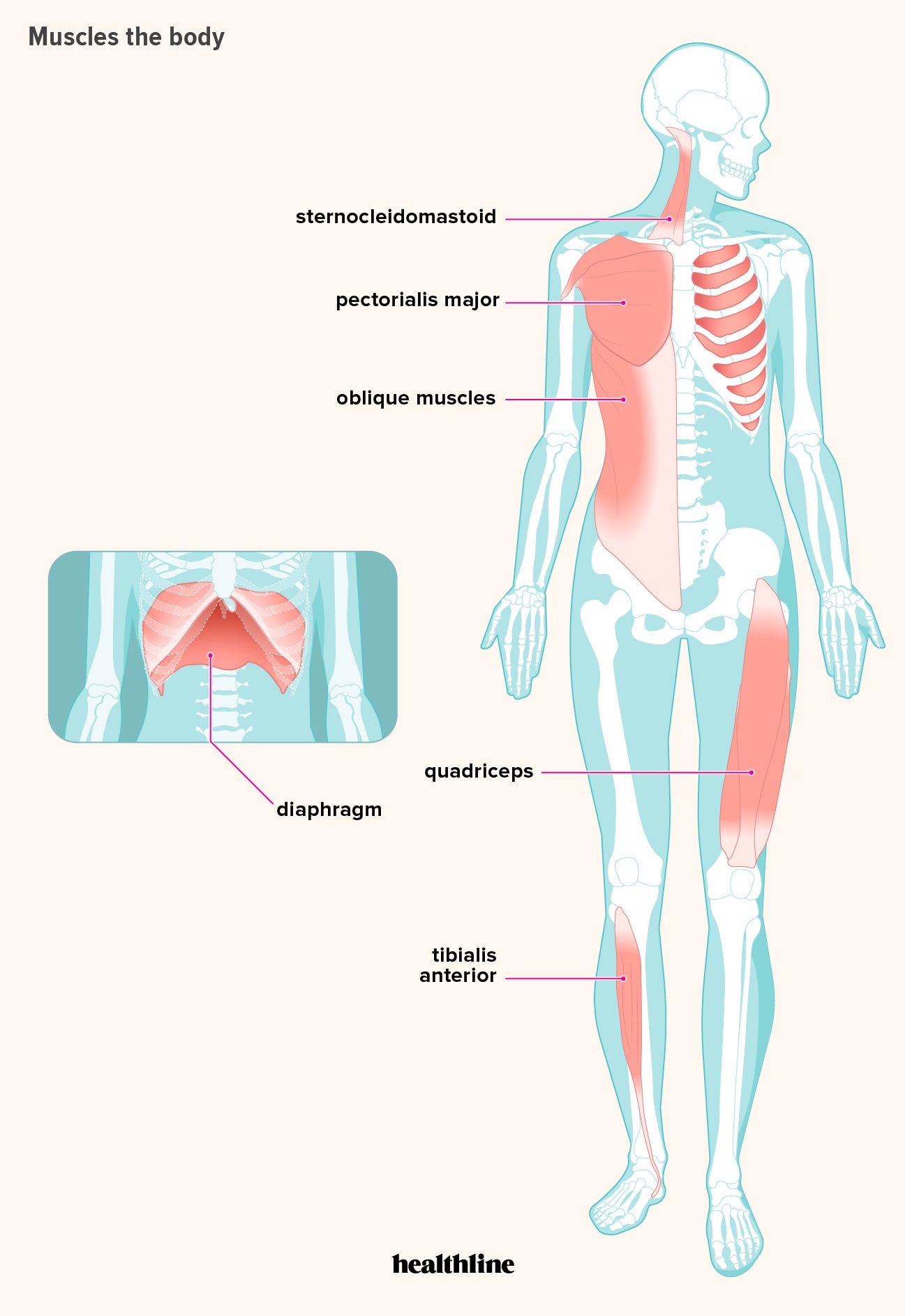
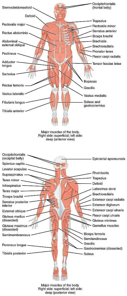
Below are two human body muscle diagrams, showing the front and back of the body.

The human muscular system is complex and has many functions in the body.

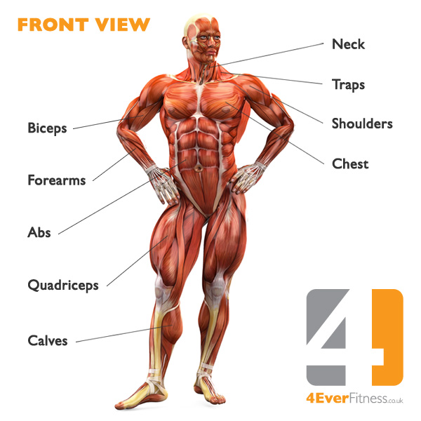
Muscle diagram, most important muscles of an athletic black man, anterior and posterior view, male body.

Located immediately below the skin) muscles of the body.

If you found any images copyrighted to yours, please contact us and we.

It permits movement of the body, maintains posture and circulates blood throughout the body.
.")
We hope this post inspired you and help you what you are looking for.

This is what happens in the body.
.")
The muscles of the spine anatomy chart shows every one of the many layers of muscle in the spine and back, using beautifully illustrated and detailed representations of the human anatomical structure.

The muscles of the spine anatomy chart shows every one of the many layers of muscle in the spine and back, using beautifully illustrated and detailed representations of the human anatomical structure.

There are approximately 640 skeletal muscles within the typical human, and almost every muscle constitutes one part of a pair of identical bilateral muscles, found on both sides, resulting in approximately 320 pairs of muscles, as presented in this article.

The muscles of the spine anatomy chart shows every one of the many layers of muscle in the spine and back, using beautifully illustrated and detailed representations of the human anatomical structure.

Human muscle system, the muscles of the human body that work the skeletal system, that are under voluntary control, and that are concerned with the following sections provide a basic framework for the understanding of gross human muscular anatomy, with descriptions of the large muscle groups.

Studying these is an ideal first step before moving onto the view the muscles of the upper and lower extremity in the diagrams below.

Gas trocsoleus (gastrocnemius and soleus muscles).

Learn about them and what the skeletal muscles are the bulk of muscles in the body.

Muscle diagrams are a great way to get an overview of all of the muscles within a body region.

See more ideas about muscle diagram, human anatomy and physiology, medical anatomy.

But, your soleus muscle in your lower leg and muscles in your back involved in maintaining posture contain mainly slow twitch muscle fibres.

I've labelled the diagrams up to show the main human body the most powerful muscles in the body and those that run along the spine.
 muscles of the body.")
Located immediately below the skin) muscles of the body.

The ear contains the smallest muscles in the body alongside the smallest bones.

Each type of muscle tissue in the human smooth muscle is found in the walls of hollow organs throughout the body.

The ear contains the smallest muscles in the body alongside the smallest bones.

Their main function is contractibility.

Posting Komentar untuk "Muscles In The Body Diagram - Solved Draw The Free Body Diagram Showing The Forces That Chegg Com"